
AWARDEE: Zhen Xu
FEDERAL FUNDING AGENCIES: National Institutes of Health, Department of Defense
“Histotripsy” doesn’t exactly roll off the tongue. If one isn’t careful when first trying to pronounce it, the word can get stuck. It might require another try – even multiple tries. In many ways, this reflects the experience of Zhen Xu, the biomedical engineer who discovered and helped develop histotripsy. Throughout her career, Xu has encountered one roadblock after another, yet she has tirelessly worked to develop the medical technique. Today, it is used in clinics around the world to treat patients with liver cancer.
Zhen Xu (Brenda Ahearn, University of Michigan)
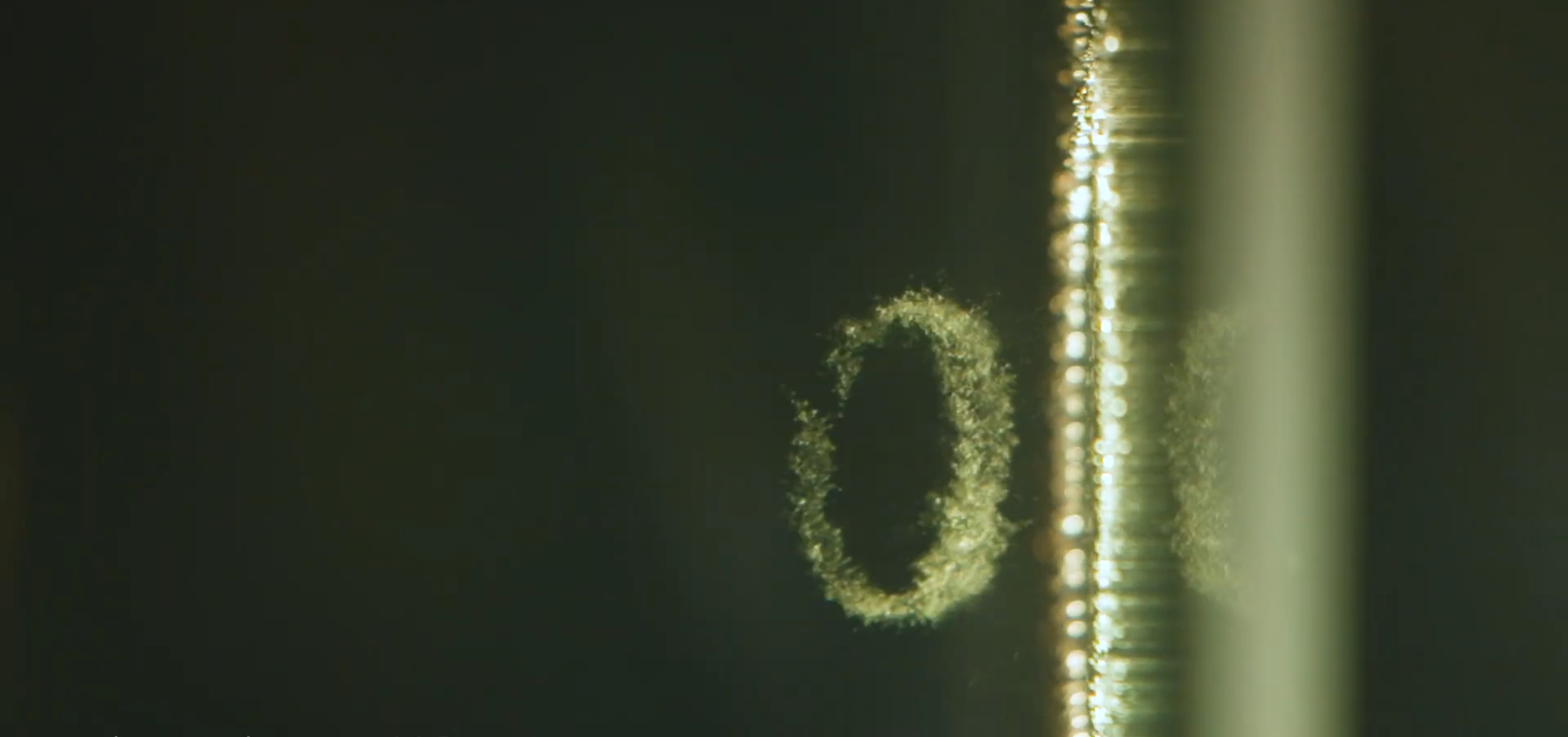
Histotripsy is most easily described as a process that liquifies tissue. On a cellular level, it works by mechanically pulling cells apart, with tiny bubbles serving as the scalpel. These bubbles expand and contract repeatedly, and eventually the motion pulls the offending tumor cells apart. The body then naturally absorbs the remaining cellular debris.
Watching the technique on a screen, histotripsy specialists can see the targeted tumor shrinking and eventually collapse. Without an incision, the surrounding healthy tissues sustain no damage.
“It’s very much like a surgery — but not invasive,” Xu says. And just like a surgery, the tumor is carved out with precise margins, but rather than a surgical tool, the histotripsy machine does it through ultrasound delivered externally. Patients can go home immediately after the treatment, and most experience minimal to no pain.
“There’s no real injury to recover from,” she continues. “It’s ultrasound-generated microbubbles that mechanically destroy tumors, and critical structures like nerves and major vessels are more resistant to histotripsy than tumors, so this is good for tumors that are near the critical structures and hard to operate on.”
Not for the Squeamish
Xu’s histotripsy journey started in 2001, when she left her native China and came to the United States for a Ph.D. in biomedical engineering at the University of Michigan.
Her parents had hoped she’d become a doctor, but Xu wanted to help people in other ways. “I was good at math and physics, and engineering was a natural fit.” Biomedical engineering proved to be the “happy marriage” of all of those interests.
“The U.S. had the best biomedical engineering graduate programs. I applied to a bunch of schools, and Michigan gave me a full fellowship.” She’s been in Ann Arbor ever since.
The serendipity began when Xu found her way to the person who became a beloved mentor: Charles Cain. Cain inspired Xu to steer into independent thought and sticky problems. At one point, he invited a guest speaker to his research group, pediatric cardiologist Achi Ludomirsky, who issued the students a challenge. Ludomirsky wanted to help very young children with congenital heart disease; they needed surgery, and tragically, not all of them were able to survive the invasive procedure. He envisioned a new way forward that would involve removing heart tissue in a non-invasive way.
“I thought he was so inspiring, and I said I wanted to take it on,” she recalls. She had no experience with ultrasound.
One of the first roadblocks was finding hearts on which to conduct her experiments. Xu turned to the yellow pages, the neighborhood phone directory that existed in nearly every household before the internet age, and located a nearby slaughterhouse that processed pork products. The owner told her to swing by early that Thursday morning. He asked if she was squeamish. Xu didn’t know the meaning of the word, but she sensed the right answer was “no.”
“I didn’t have any sense of normal,” she says. “I just knew I got a heart, and it cost $5.” So, every Thursday, she’d return with her $5 and dig through dumpsters to get the tissue she needed. Squeamish? No way.
Quiet, Please
The experiments themselves presented more challenges: They kept failing. “When you do ultrasound therapy, you do bursts [of sound waves] and repeat them in frequencies,” Xu says. The ultrasound machine she was using was high-powered and produced unusually long pulses. She kept trying different repeating frequencies, to the point where she hit the audible range. Still, she wasn’t making headway. To add insult to injury, her lab mates started complaining about the noise.
“I thought, if nothing is working anyway, try a bigger hammer,” she recalls. Xu decided to amplify even further. But she needed to do it without driving her fellow researchers mad. She found an old and powerful amplifier and turned the knob all the way up. At the same time, she employed a superfast, microsecond pulse sequence to keep the noise down. This was a new combination … and it caused something unexpected to happen. Xu saw what looked like tissue disintegrating before her eyes.
“I ran over to Charles [Cain] and said ‘something special just happened,’” she remembers. “He said ‘repeat it.’”
And there was something else. “I had to tell him I just broke the most expensive machine we had.”
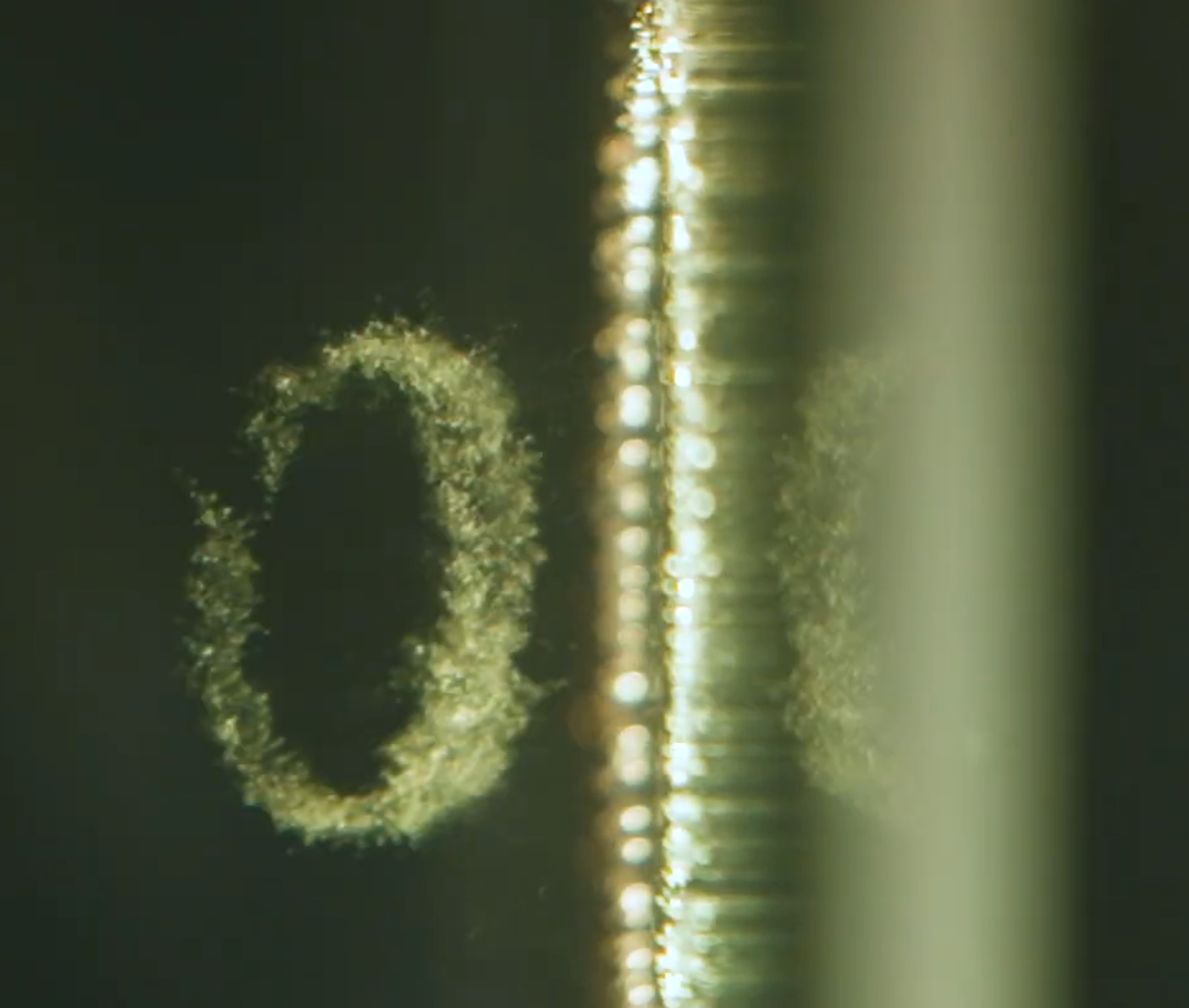
Histotripsy bubbles (Zhen Xu)
The Pivot Point
With this unexpected breakthrough, Xu pivoted to focus her doctoral training on the development of histrotripsy. The name “histotripsy” was intentional: “histo” is Latin for “tissue,” and “tripsy” indicates a “mechanical breakdown.”
Working with Cain and a multidisciplinary group of colleagues, Xu put out the first publication on the technique in 2004. At the time, it was met with some skepticism in the community, she recalls. The following year, the team worked to develop their own device, building on Xu’s discovery, with key contributions from lab mate and fellow engineer Tim Hall. 2005 was also the year she completed her doctorate. A few years after that, the company HistoSonics launched to help move the technology forward.
The National Institutes of Health was a key supporter starting in the early years of Xu’s work. In time, the Department of Defense also became a funder.
“The federal government was incredibly important to support this work over the many years of failures before monumental success,” says Karen Thole, who served as the engineering dean at the University of Michigan when the technology was making its way from the lab to the clinic. “I got to witness it being birthed,” she recalls. “I started watching the numbers of patients. The impacts were amazing.”
Without this federal funding, “I don’t think we would have been able to do this,” Xu agrees, “especially since in the beginning, it was considered impossible to do.” The U.S. government, she adds, has been a “driving force behind innovative technologies.”
From Bench to Bedside
By 2016, it was time to channel the years of effort into a clinical trial with actual patients. The trial focused on prostate tissue. While Xu and her colleagues were hugely enthusiastic about the potential, and the trial proved safe for the patients, it did not liquefy the tissue as they’d hoped. It was another blow on the years-long histotripsy roller coaster. But Xu persevered. She and her collaborators shifted more of their focus to liver cancer after seeing strong pre-clinical results. A couple of years later, they saw more promising results in a liver cancer clinical trial in Spain. This led to another, larger clinical trial called HOPE4LIVER. It was a success.
In 2023, more than two decades after Xu’s discovery, the U.S. Food and Drug Administration approved histotripsy as a treatment for liver cancer. It has now been used in thousands of patients and opened up a whole new field of work.
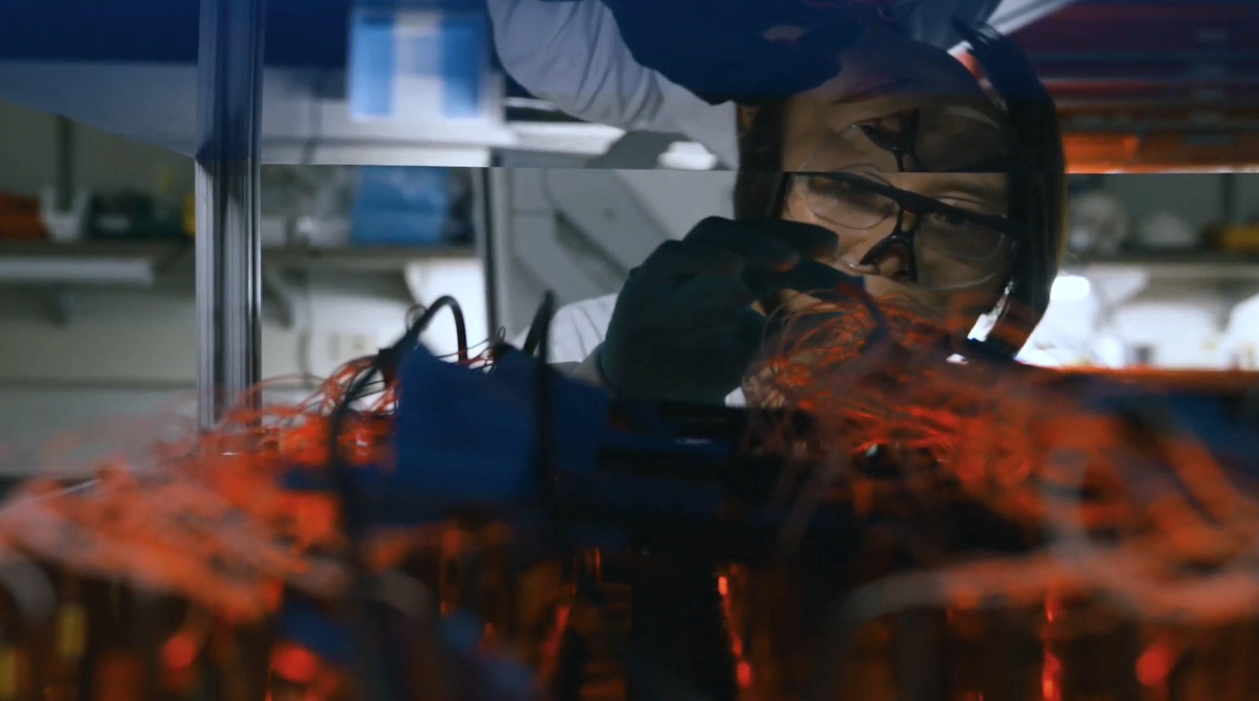
Zhen Xu (522 Productions)
While Xu’s focus on histotripsy has been consistent, the path has been anything but. “This is a true grit story,” says Thole, now at Penn State. It is rare that a scientist will make her grand discovery before she even earns her Ph.D. Xu has spent her entire career building the knowledge and tools to further the field of histotripsy and bring it to patients. Yet, science doesn’t happen in a vacuum, and she is quick to acknowledge the many colleagues at the University of Michigan, HistoSonics and elsewhere who have helped bring the vision to life. The work is far from finished.
Promising clinical trials are underway for both kidney and pancreatic cancer patients. Furthermore, the company that launched out of the histotripsy work, HistoSonics, is now backed by companies associated with household-name investors like Jeff Bezos and Peter Thiel. At press time, it is valued at $3.75 billion and boasts hundreds of employees. Xu remains a scientific advisor there. And on September 10, Xu and her colleagues launched the University of Michigan Histotripsy Center. “The idea is to set up workstations and equipment and train personnel to open up the work I’ve been doing to a new community, so [researchers] can try this on new diseases,” she says. “We also have a travel histotripsy machine that we can send to different locations to expand the scope of research, especially in areas where funding can be tight.” She adds that these resources will be free for researchers to use.
Xu often thinks of the patients she’s met who have undergone histotripsy. Many of them have had to try multiple painful or invasive treatments to keep their liver cancer at bay. What they tell her, again and again, is that because the treatment is noninvasive and requires a minimal recovery time, it gives them more time to spend with their families and do things that are truly meaningful to them – a better quality of life. “That,” Xu says, “is what is most rewarding.”
By Erin Heath