

AWARDEES: David Deamer, Mark Akeson, and Daniel Branton
FEDERAL FUNDING AGENCIES: National Institutes of Health, National Science Foundation, Defense Advanced Research Projects Agency, National Aeronautics and Space Administration
David Deamer was driving along a forested road in Oregon in 1989, thinking about his research at the University of California, Davis. Deamer was investigating how to improve the understanding of DNA, the key chemical in the genes of all living organisms. DNA is a long, flexible string-link material formed by the linkage of each of the four smaller chemicals called bases that are abbreviated as A, C, G, and T. These bases are chemically attached to each other in many different sequences such as … CGATTCACCCATATG... Determining the order or sequence of bases in DNA is enormously important; just as the story in a novel is carried by the sequence in which any of the 26 letters in the English alphabet appears on a page, so too does the sequence to which A, C, G, and T bases are linked to each other. The order carries the blueprint, or genes, of all living organisms.
Deamer recalled that the four bases of DNA are all slightly different in size, and that realization unlocked a novel idea about how he could determine the sequence of bases in DNA. He immediately pulled to the side of the road and scribbled down the original concept that would eventually become nanopore sequencing, a method that has enabled some of the most significant advances in the field of genomics. But the road to success took years of research, persistence, and a bit of serendipitous fortune to prove many skeptics wrong.

David Deamer’s original notebook sketch, 1989 (David Deamer)
Deamer wrote in his notebook that day, “DNA will be driven through a small channel…the channel will be carrying a current. As each base passes through, a change in the current will occur. Because the bases are of different sizes, the current change will be proportional, thereby providing an indication of which base it is.” He then continued his drive. For two years, the sketch was just an idea scribbled in his notebook.
Federal Investment in DNA Sequencing Technology
In the mid-to-late 1980s, discussions were just starting to ramp up about the feasibility of sequencing human DNA. Prior to this time period, DNA sequencing was quite limited and depended on slow, tedious methodologies. By 1990, the Human Genome Project, a large international collaborative project to generate the sequence of all 3.2 billion letters in the human genome, was launched.
Early on, the National Center for Human Genome Research (NCHGR) at the National Institutes of Health (NIH), which in 1997 became the National Human Genome Research Institute, was focused on both sequencing the human genome and advancing the technology involved with sequencing. The latter goal meant expanding granting practices to identify and fund high-risk but high-payoff proposals. Nanopore sequencing was one of these high-risk but high-payoff proposals. Until NCHGR was created in 1992, this type of granting practice was entirely outside the norm at NIH.
Two years before the human genome project reached completion, the federal government had invested $500 million into sequencing the first human genome by 2001 and there was great interest in reducing the sequencing cost. The Advanced Sequencing Technology Program (ASTP) at NCHGR focused on funding proposals to improve sequencing technology, which reduced sequencing costs to $1,000 per human genome. Providing funding to develop a dramatic improvement in technology was inherently risky but ultimately achieved a very high payoff. The team in this story received early funding from the National Science Foundation and DARPA as well as continued funding from NASA and NIH’s sequencing programs.

Daniel Branton
How Two Team Members Met
In the 1960s, Daniel Branton was an assistant professor in the Botany department at UC Berkeley. Deamer was a UC Berkeley postdoc working one floor above Branton’s lab. At the time, Branton was exploring a new technique called freeze-fracturing to understand how proteins interact with cell membranes. Most cell biologists did not accept Branton’s interpretation of the microscope images, which he believed showed the freeze-fracturing process to split biological membranes into two sheets. Deamer was intrigued by Branton’s interpretation and joined him in the lab. The results of this work were published as a cover article in Science and significantly advanced our understanding of the structures of cell membranes. It was also the beginning of a lasting friendship that later initiated their research on nanopore sequencing.

David Deamer
Fast forward to 1991. Branton, now a member of the biology faculty at Harvard University, was invited by Deamer to visit UC Davis and give a series of lectures. At the time of his visit, Branton had been contemplating the current methods for DNA sequencing. He thought to himself, “There must be a better way of doing this.” Could the individual bases be identified by measuring the force required to pull each base in a single strand of DNA through the interface between water and air? During their discussions, Deamer shared his idea, scribbled in a notebook from a couple of years earlier, which outlined using an electrical voltage to pull a strand of DNA through a small pore. Branton thought Deamer’s idea was much better than his own and they began collaborating on the nanopore sequencing concept.

Illustration of a nanopore
Serendipity and Skepticism
After they decided to explore what Deamer had fortuitously sketched, they moved forward to identify the best channel, or “pore,” through which to move the DNA. They identified a protein called haemolysin as possibly having a pore wide enough to accommodate a single strand of DNA. In 1993, Deamer met with John Kasianowicz at the National Institute of Standards and Technology (NIST) to test a haemolysin nanopore. “Nanopore” refers to the size of the pore, which is approximately 2 nanometers in diameter (for comparison, a sheet of paper is about 100,000 nanometers thick!). In Deamer’s notebook, he had written, “the thickness of the membrane must be very thin…the channel must be of the dimensions of DNA in cross-section, approx. 1-2 nanometers.” The early experiments Deamer and Kasianowicz performed showed positive confirmation of Deamer’s sketched idea — that a voltage applied across the pore could draw a single strand of DNA through the channel. This finding was a pivotal moment in their work.
The National Science Foundation (NSF) awarded the team $50,000 in 1994 to help sustain the team’s experiments for a year, and then it was time to publish what they had learned. But both Nature and Science rejected their submission. “Nobody could believe it,” Deamer recalls. The scientific community was still skeptical of their findings. Thanks in part to Branton being a member of the National Academy of Sciences, their paper was accepted and published in the Proceedings of the National Academy of Sciences (PNAS) in 1996. The paper would turn out to be a seminal, decisive contribution cited thousands of times.
Concurrently in 1994, Deamer had moved from UC Davis to UC Santa Cruz, and around the same time, he and Branton decided to pursue a patent for their idea. Having a patent would be important if their nanopore idea was going to be transformed into a commercial product. Translating a research idea into something usable for scientists, clinics, or hospitals is a huge undertaking, requiring a large and risky financial commitment from outside investors. Their work was not ready for that next step quite yet, but having the patent would prepare them for what they hoped would be future opportunities to catalyze their work.
Deamer and Branton submitted their idea to Harvard’s patent office and quickly learned another Harvard faculty member, George Church, a well-known geneticist, had a similar idea that involved driving double-stranded DNA through a nanoscopic channel. The three professors – Branton, Church, and Deamer – decided they would all be listed as inventors. This ended up being very fortuitous because Harvard’s patent office expressed skepticism about pursuing a patent for such a risky idea as nanopore sequencing. Church gave their idea more credibility by arguing that if three professors at different institutions had a similar idea, the idea must surely have some merit. Church’s argument helped convince the patent office to proceed with an application.

Mark Akeson
Nanopore sequencing continued gaining momentum: the team received NIH and DARPA grants in 1997 and were awarded patent rights in 1998. And in 1996, the PNAS paper convinced a junior colleague, Mark Akeson, to join the collaboration. Akeson, who had previously trained with Deamer at UC Davis, was a post-doctoral fellow at NIH in Bethesda, Maryland. When Deamer initiated one of his early experiments with Kasianowicz, he visited Akeson, who recalls driving Deamer to NIST thinking (along with many others at the time) “This idea is nuts!” But once Akeson read the pivotal PNAS paper, he decided to leave his stable position at NIH and moved across the country to rejoin Deamer. It was a risky move, but for Akeson the chance to work on a revolutionary sequencing concept won the day.
By 1997, Akeson, Branton, and Deamer (along with collaborators Kasianowicz and staff scientist Eric Brandin at Harvard University) took their work to the next level. Now that they had a working pore, they needed to test if it could distinguish between the A, G, T, and C bases. If it couldn’t make any distinction whatsoever, the nanopore sequencing concept might never work. In 1999, research led by Akeson demonstrated that the nanopore could distinguish between stretches of C versus A bases composing individual RNA molecules. Akeson and Deamer, along with graduate students Wenonah Vercoutere and Stephen Winters-Hilt, subsequently published a paper in 2001 that showed individual A-T or G-C base pairs could be distinguished at the end of single DNA molecules suspended in a nanopore. This level of nanoscopic precision “finally convinced a bunch of skeptical people that we knew what we were doing,” Deamer recalls.
Controlling the DNA in the Nanopore Sensor
The collaborators had shown that long segments of two different bases could be distinguished as they passed through a protein nanopore, but sequencing would require distinguishing between individual bases, not segments, within each DNA strand. This was not possible in 1999 because strands of DNA and RNA (driven by a strong electric field) rocketed through the nanopore at roughly one microsecond per base (one-millionth of a second) – too fast to read single bases. One possible solution was to use a processive enzyme (a ‘molecular motor’) to reduce the translocation speed by approximately a thousand-fold. In the mid-2000s, Akeson’s group at UC Santa Cruz and a group led by Hagan Bayley in Oxford, UK, found that several DNA ‘motors’ could bind and slow DNA movement through nanopores, but only for one or two bases before slipping away. According to Akeson, “This result was frustrating and concerning, because the strength of the electric force acting on DNA in the nanopore was unknown, so it was a real possibility that no molecular motor could bind DNA strongly enough to allow long strings of bases to be sequenced.”
Fortunately, their perseverance paid off. In 2010, the UC Santa Cruz group was able to secure a molecular motor to translocate DNA strands back and forth through the alpha-haemolysin nanopore at the length of one base in DNA. More than 500 DNA molecules in series were moved one-by-one in precise order through the nanopore under robotic control.
Subsequent Work and Continuing Impact
In 2007, Deamer, Akeson, Branton, and his Harvard collaborator, physicist Jene Golovchenko, were approached by Oxford Nanopore Technologies (ONT) to license patents related to nanopore sequencing with the hope of commercializing the technology. All four gave permission, and within seven years, ONT developed and brought to market the MinION, a $1,000 pocket-sized sequencing device. Unlike traditional sequencing approaches, which need time-consuming computer analysis after sequencing is completed, nanopore sequencing produces direct, real-time sequence reads as soon as the sequencing process begins. Furthermore, nanopore sequencing can read longer strands of DNA than most other devices and has been used virtually anywhere, including the top of an arctic iceberg, a remote settlement in Africa where electrical power is unreliable, and on board the International Space Station.
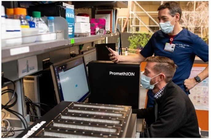
Euah Ashley and John Gorzynski performing nanopore sequencing at Stanford University Hospital, 2022

Astronaut Kate Rubin sequencing DNA with a MinION on the International Space Station (NASA)
In 2022, this combination of speed and read length helped deliver human genomes for intensive care patients at Stanford University Hospital. One of the sequencing tasks set a speed record of five hours for a human genome, and another was credited for saving a young boy’s life by identifying a genetic mutation causing heart disease.
The work to develop nanopore sequencing into what it is today took over 30 years of commitment to pursuing basic scientific understandings. This eventually led to the advancement of a commercially viable product. Nanopore sequencing has been widely used for pathogen analysis in outbreak surveillance of infectious diseases such as tuberculosis, Ebola, Zika, and dengue fever, among others. Many of the sequenced COVID-19 genomes globally used nanopore sequencing, making it crucial in the fight to end the pandemic. Additional applications of the technology continue to be unearthed, and it was all possible thanks to an idea scribbled in a notebook.
By Meredith Asbury